-
-
CL Solutions
Cathodoluminescence solutions that reveal fundamental properties of matter
-
Cryo Solutions
Integrated and automated solutions to u nlock the power of cryo - ET workflow -
FAST IMAGING
Fast EM solutions for reliable and high throughput electron microscopy
-
CLEM Solutions
Integrated correlative microscopy solutions that combine the power of fluorescence and electron microscopy
-
CL Solutions
- Contact
- All resources
-
CL Solutions
Cathodoluminescence solutions that reveal fundamental properties of matter
-
Cryo Solutions
Integrated and automated solutions to u nlock the power of cryo - ET workflow -
FAST IMAGING
Fast EM solutions for reliable and high throughput electron microscopy
-
CLEM Solutions
Integrated correlative microscopy solutions that combine the power of fluorescence and electron microscopy
Cryo Solutions
METEOR: frequently asked questions
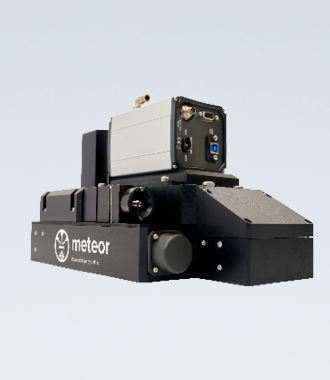
Learn more about the specifications of our integrated top-down cryo-CLEM imaging system.
General
-
Is METEOR compatible with my current FIB/SEM?
METEOR is designed to be retrofitted to any existing FIB/SEMs. If you have questions regarding your specific configuration, please contact us by filling out the enquiry form and one of our sales representatives will contact you.
-
What is the price of METEOR?
If you want a quotation of METEOR please contact us by filling out the enquiry form and one of our sales representatives will contact you.
Hardware
-
What are the specifications of available lenses?
The following options are available*:
Olympus Fluorite objective
10x (WD 11.0 mm, NA 0.30)
20x (WD 3.1 mm, NA 0.45)
50x (WD 1.0 mm, NA 0.80)
100x (WD 1.0 mm, NA 0.90)
Olympus Apochromat objective
50x (WD 0.35 mm, NA 0.95)
100x (WD 0.35 mm, NA 0.95)
Semi-Apochromat objective50x (WD 10.6 mm, NA 0.5)
*Depending on shuttle and sample stage configuration the choice of objectives can be limited due to space restrictions.
-
Is the objective vacuum and cryo compatible?
The objectives are compatible with high vacuum environments. Our results show that there is no out-gassing from the objective (or METEOR itself). During operation, the objective is in vacuum and is not physically in touch with the sample, which is actively cooled. It neither poses a heat load to the sample, nor is it cooled by cryo stage.
-
How long does it take to install the system?
METEOR can be installed within a day. After the installation we take one more day to test all functionalities, align the system and evaluate the vacuum performance of the FIB/SEM.
-
Is the system plug-and-play? Can the filters and objective be exchanged?
The filters in the filter wheel and the objective can easily be replaced by the user if preferred. Please contact us for alternative camera options.
-
Is METEOR retractable?
Yes, the objective of METEOR is fully retractable to a position above the pole piece allowing the user to take full advantage of the stage movements.
-
Are you still able to have GIS needles installed together with METEOR?
Yes, the current version of METEOR only occupies two GIS ports leaving the other ports available for GIS needles. At our testing site we had a GIS needle available for platinum coating.
-
What is the angle between the objective and the sample?
The objective as a 52° angle with respect to the SEM polepiece (the same angle as the FIB column). The objective needs to be perpendicular to the sample during FLM imaging. The stage of the FIB/SEM can be tilted and rotated in such a way that the sample is perpendicular to the objective.
Software
-
How is the overlay performed in METEOR?
How the overlay is performed is up to the user, this can for example be done by using fiducial markers or other available methods. Upon installation the system will be calibrated and the relative movement from SEM and FIB imaging to FLM imaging by METEOR will be determined. This calibration allows the researcher to seamlessly switch from FLM to FIB/SEM imaging and back.
-
Does your software communicate with any of the already existing software interfaces for FIB/SEM?
The software communicates with the FIB/SEM through an XTlib adapter. In this way the stage of the FIB/SEM can be controlled to allow for easy sample navigation. ODEMIS, a user-friendly open-source acquisition software, is installed together with METEOR. Users can implement their own scripts in Python to automate routine processes, e.g. camera exposure time optimisation.
-
Once the sample is loaded, do you operate all actions from the Delmic control PC?
All the normal actions, like FIB milling, SEM imaging or GIS coating are controlled from the FIB/SEM control PC. Once the user decides to use METEOR actions such as moving the FIB/SEM stage, focussing the objective and taking FLM images are done through the Delmic control PC.
-
Are the output images of METEOR compatible with downstream correlation software or image processing software?
METEOR images are saved in the OME-TIFF format that can be processed using various image processing software programs like ImageJ (Fiji) and Huygens deconvolution from SVI. Correlation software like the 3D correlation toolbox can also be used in combination with METEOR data.
Imaging
-
How sensitive is METEOR towards fluorescence detection?
Like any wide field fluorescence microscopes, the sensitivity of the METEOR depends on the numerical aperture of the objective and the quantum efficiency of the camera chosen by the customer.
Our results show that METEOR is suitable for cryo biological samples expressing eGFP and nanodiamonds as small as 140 nm in diametre.
Please contact us for more information about our standard camera offered with METEOR, and alternative choices.
-
Does milling, platinum coating or electron beam imaging have any effect on the intensity of fluorescence emission?
Platinum coating and the low resolution SEM imaging that are being used in the cryo-ET workflow did not have a significant effect on the fluorescent signal of the sample in our experimentts. Milling away material using the FIB results in a thinner sample, which will result in a dimmer image due to fewer photons present in a smaller volume. However, the fluorescent region still present in the lamella is visible with a higher contrast.
-
Could the incident light cause devitrification of the sample?
We have not seen any sign of devitrification in the tomograms of the samples imaged using METEOR. It is known that high power laser sources that are for example used in super resolution applications can devitrify samples1. This is the reason we decided on a LED light source that provides more subtle sample illumination that is not expected to devitrify the sample even at long exposure times. According to our experiments the LED light source is brilliant enough to image biological samples expressing eGFP.
Reference:
1. Tuijtel, M. W., Koster, A. J., Jakobs, S., Faas, F. G. A. & Sharp, T. H. Correlative cryo super-resolution light and electron microscopy on mammalian cells using fluorescent proteins. Scientific Reports 9, 1–11 (2019).

Didn't find an answer?
Fill in the form with your question, and our experts will get back to you soon!
