-
Life Sciences
.png)
Our solutions
-
Materials Analysis
.png)
Our solutions
Techniques
Applications
- Why Delmic?
-
Insights
.png)
Insights
-
Company
.png)
Company
Marine microbiology
Studying marine microorganisms with integrated CLEM

Dr. Sten Littman is a scientist working at the department of biogeochemistry in MPI for Marine Microbiology in Bremen. His work is dedicated to imaging and microanalysis of micro-organisms using Scanning Electron Microscopy (SEM) and energy-dispersive X-ray spectroscopy and on single-cell environmental microbiology.
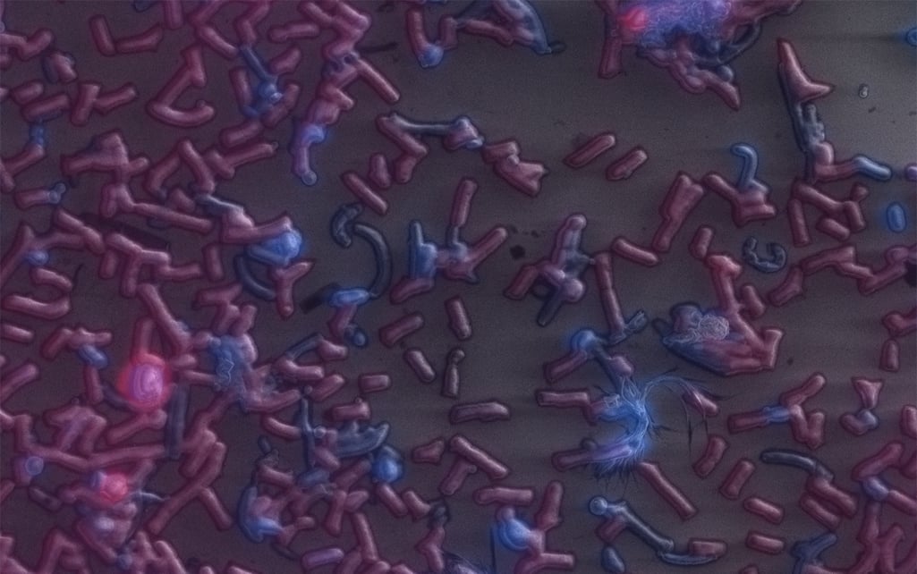
Studying microorganisms (or microbes), which are found in the ocean waters, is a fascinating process that can reveal the hidden secrets about ocean chemistry, biology and climate. Marine microorganisms are exceedingly small, diverse in their forms and distributed across the ocean, which makes it so challenging to analyze them. For more than four years he has been using the SECOM, integrated correlative light and electron microscope, for his research. This system combines fluorescence and electron microscopy allowing to investigate the structure of the specific regions.
This way of studying marine microorganisms is extremely convenient because of one special quality of certain microorganisms: autofluorescence of the natural emission of light. Because of this, fluorescence microscopy can be a great tool to identify the microbes and image their physiological and metabolic state. Simultaneously, electron microscopy is used to get the high-resolution structural information.
According to Dr. Littman, the SECOM helps him to gain more insight and advance in his research:
It gives us the possibility to identify different types of bacteria in environmental samples by the hybridization of these cells with specific HRP probes, after which we image them with the SEM and perform elemental analysis, including elemental analysis with EDX.


